

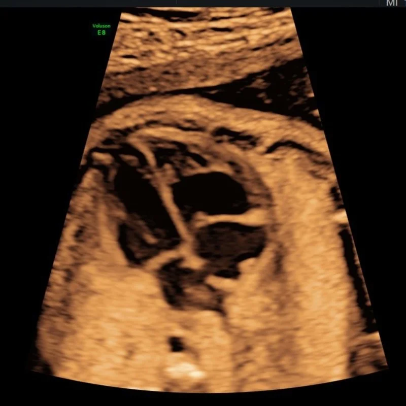
Morphology Scans
From 21 weeks
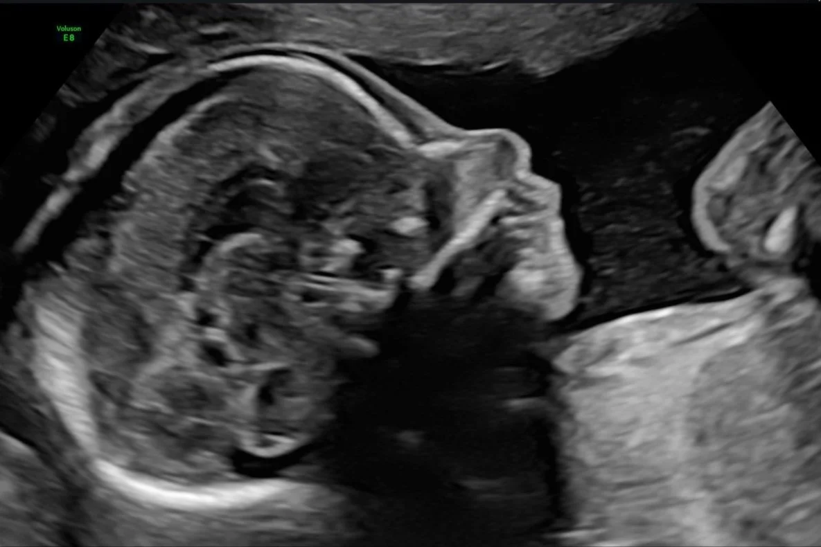
Morphology Scans
A routine Ultrasound examination is offered to most women at 19-22 weeks. This scan is often referred to as a Morphology or 20 week ultrasound.
The purpose of the Morphology examination is to:
Confirm the fetus is alive
Identify multiple pregnancy if no earlier scans were performed.
Confirm Estimted due date (EDD) if no earlier scans were performed.
Estimate and assess the fetus’ gestational age and early fetal growth by measuring the fetus’ head, abdomen, femur and humerus.
Perform a detailed assessment of the developmental anatomy of the fetus’ head, brain, face, lips, palate, heart, diaphragm, lungs, kidneys, abdominal wall, bladder, spine, skin line, arms, hands, legs and feet.
Fetal gender can be performed on request.
Position of the placenta and umbilical cord insertions
Length of the cervix
Amniotic fluid
Pelvic anatomy i.e. fibroids and ovarian cysts
Measurement of the uterine arteries
The Morphology Scan is expected to detect about 60-80% of fetal malformations. However, not all abnormalities can be detected despite diagnostic views.
Visualisation of the fetus’ anatomy maybe limited by the fetus’ position and movement. Maternal BMI is also a contributing factor as increased body fat significantly reduces the quality of the images.
During the scan, every attempt is made to move the fetus to obtain diagnostic images. Occasionally, we may have to ask you to return at a later date to complete the examination when the fetus is bigger and/or in a different, hopefully better position.
Some congenital heart defects, bone growth and bowel obstructions are progressive and unable to be detected at this stage. Also, biomechanical and some chromosomal abnormalities and cerebral palsy cannot be detected.
At Trinity Imaging Ultrasound for women, we preferably like to perform the Morphology scan at 21 weeks to obtain the most optimal diagnostic images.

-
To acquire the best diagnostic images possible, the examination is routinely performed Transabdominally, followed by a Transvaginal ultrasound (always with your written consent first).
-
In most cases the examination will be performed transabdominally, but there are some situations where a Transvaginal ultrasound maybe necessary. This improves the assessment of the cervix, placenta and obtain better details of the fetus. In turn this can improve accuracy if the diagnosis.
Transabdominal and transvaginal ultrasound examinations are safe at all stages of pregnancy.
-
At this stage, the fetus size makes it difficult to measure the entire length accurately. However, we can calculate the weight using several measurements. While we cannot predict the fetus size at delivery based on this scan, we can assess whether the fetus current size is appropriate for your stage of pregnancy.
-
Diagnosing Down syndrome through ultrasound is not possible, as it is caused by a chromosomal abnormality. Even during a 19-week scan, around 40-50% of babies with Down syndrome may appear completely typical. However, in some cases, certain markers may be identified during the scan, leading to increased suspicion of a chromosomal abnormality in your fetus. If this occurs, the findings and next steps will be discussed in detail with you and your doctor.
-
While a morphology scan can provide a wealth of details, it's important to understand that a normal scan cannot offer a 100% guarantee of a normal fetus. Not all parts of the fetus will be clearly visible on this scan, and even with an experienced sonographer and advanced ultrasound equipment, some abnormalities may not be detected. Research consistently indicates that morphology scans are unable to detect all fetal abnormalities. With approximately half of major anomalies being visible and the other half not. Certain issues, such as heart defects, bone growth problems, and bowel obstructions, may not be evident until later in the pregnancy. Additionally, in larger women, the diagnostic accuracy is further reduced due to the impact of increased body fat on the quality of the images obtained.
If you have specific concerns related to family history or elevated risk identified during first trimester screening, it's crucial to communicate this information to your Sonographer at the beginning of the scan.
-
It's important to recognize that a fetal anatomy survey may not detect all fetal problems, and the accuracy of the scan can be influenced by factors such as the fetus' position, gestation, and the mother's build. For women with higher body fat, the diagnostic accuracy may be reduced.
While ultrasound is highly effective in detecting conditions like neural tube defects (such as anencephaly and spina bifida) with a detection rate of 90%, the detection rates for conditions like cardiac defects are notably lower (25-50%). Even with the best expertise, it's simply not possible to identify every fetal problem. The main objectives of the fetal anatomy survey are to verify the accuracy of pregnancy dating, assess the position of the placenta, and potentially recognize significant fetal structural issues that could impact the location, timing, or method of delivery. It's important to understand that no prenatal ultrasound examination can identify all fetal structural problems.
If your pregnancy carries a specific risk of a particular fetal abnormality, such as a family history of spina bifida, please inform us. It's crucial to note that ultrasound cannot detect conditions like cerebral palsy or autism. Furthermore, an unremarkable fetal anatomy survey does not guarantee that your baby will be free of any imperfections.

-
At the time of the ultrasound scan, it's important to note that 96% of fetus’ are found to be structurally normal. Approximately 2% of fetus’ may have a major structural problem, such as a heart defect or spina bifida, while an additional 2% may have a minor issue, such as a facial cleft lip or distension of the collecting system of the kidney.
If a problem is detected during the ultrasound scan, we will promptly inform you during the appointment. Additionally, we will communicate the findings to your doctor, engage in a discussion with them, and make arrangements for further management as necessary.
-
The ultrasound procedure typically lasts around 60 minutes. However, the duration may vary depending on the specific reason for the examination and the complexity of the individual case.
-
If possible we recommend wearing comfortable, loose-fitting clothing that provides easy access to the area being imaged. Two-piece clothing, with separate upper and lower garments, is preferable.
Additionally, please ensure that you empty your bladder 1 hour before the procedure and then drink 2 glasses (600ml) of water, holding it without emptying your bladder again. Please note that your appointment might be delayed if your bladder is not adequately full.
-
Having some urine in your bladder can be beneficial for outlining the cervix and visualizing the relationship of the placenta with the lower uterus during imaging. It also helps to elevate the uterus from the pelvis into the abdomen, facilitating better visualization of the fetus.
You don't need to have a large amount of urine, and simply drinking a glass of water 30-60 minutes before your scheduled appointment is sufficient.
-
We welcome one adult support person, whether it be your partner or a family member, to accompany you during your Diagnostic scan. However, for specific reasons, we have implemented policies that prohibit children from attending Diagnostic ultrasound appointments. While we understand that this may be disappointing for families, it is essential to maintain a focused environment. Thus allowing the Sonographer to be able to concentrate during the scan whilst taking the necessary images and measurements. Occasionally, our sonographer may need to discuss unfortunate news found during your appointment and so this must be taken into consideration. The presence of children can introduce distractions that may impact the quality of the examination.
-
At Trinity Imaging, we provide our patients with their images to an app on your phone called Tricify. For further details please ask our receptionist at your appointment.

All our scans are performed in our premium-furnished luxury viewing room specially designed for you.
We are all looking forward to getting to know you, your baby and family along your pregnancy journey.
Our Morphology Scans take up to 50 minutes and include:
2 x 3D thermal printed hard copy images
All pictures taken on the day straight to your mobile phone