

Nuchal Translucency Scans
From 13 weeks

Nuchal Translucency Scans
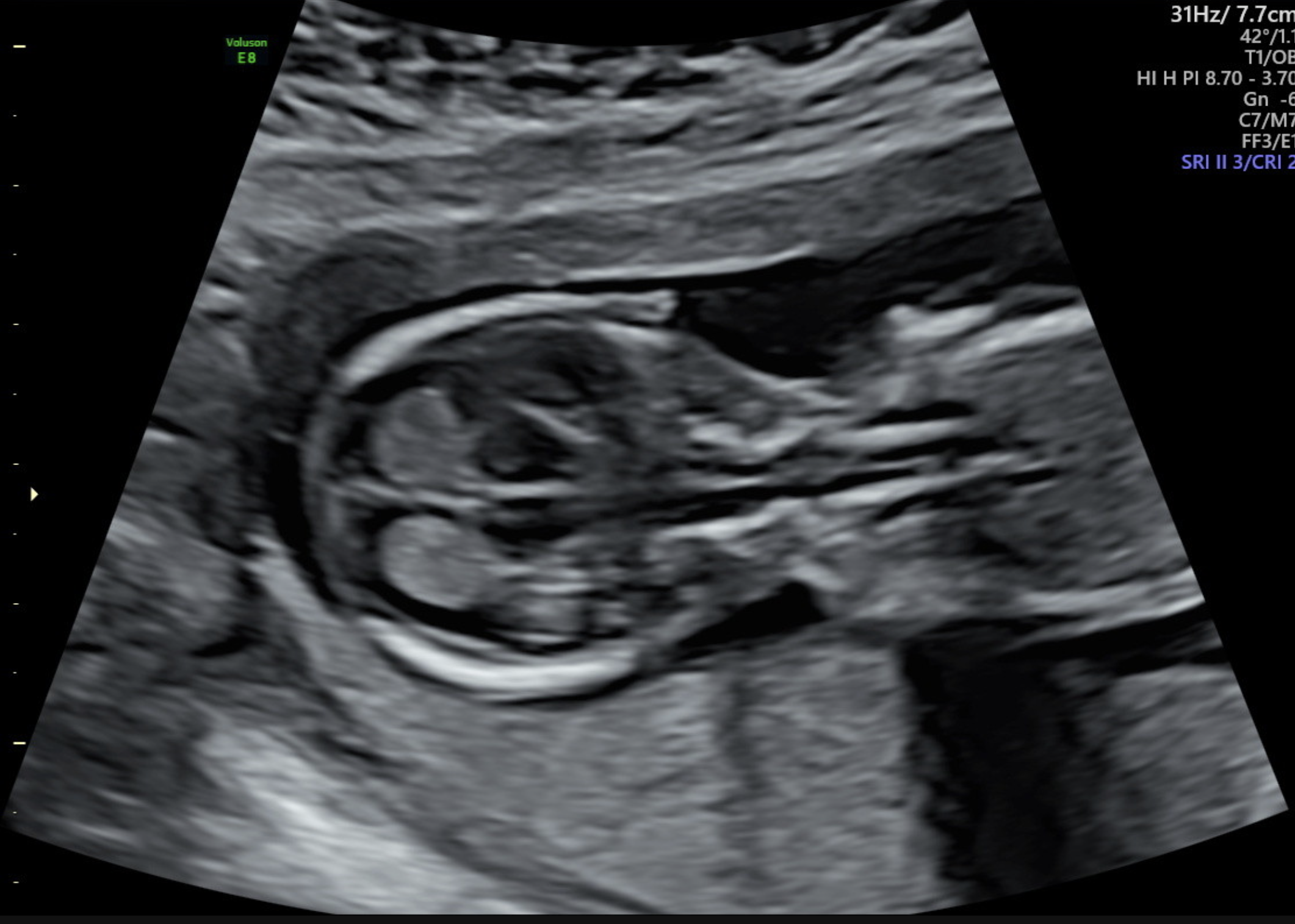
Nuchal Translucency is the Sonographic appearance of the normal fluid space behind the head and neck of the fetus. This fluid is visualised in the first trimester of pregnancy.
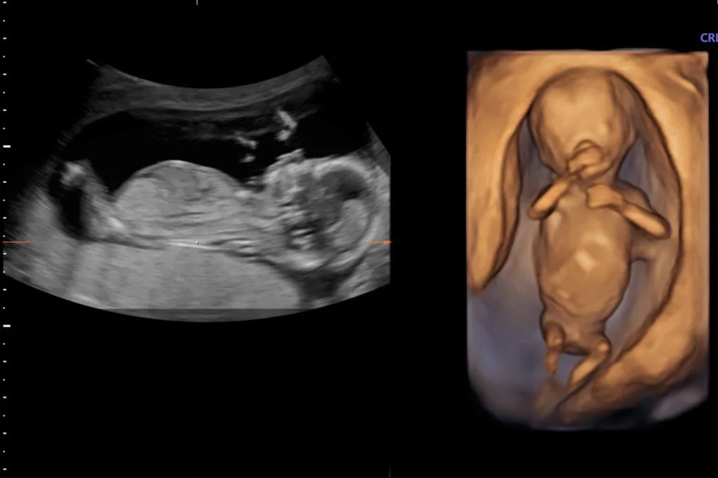
A Nuchal Translucency Scan is also a first trimester screening ultrasound. The examination offers assessment for chromosomal anomalies. The test suggests which pregnancies are at a higher risk of abnormalities and may need further investigation.
The extra fluid and Nuchal Translucency measurement tends to be thicker (>3mm) in a baby that may be associated with chromosomal abnormalities. It can be compared with what is expected for a baby of the same size (Nuchal Translucency normal range).
Other non-chromosomal conditions, such as neural tube defects, limb abnormalities and some congenital heart disease may also be detected at this stage of pregnancy.
The Ultrasound is a very time sensitive scan and the optimum time for performing this examination is between 12 weeks and 13 weeks 6 days as many cranial anatomical features which are being assessed won’t be detected until then.
At Trinity Imaging for women, we offer the first trimester ultrasound at 13 weeks of your pregnancy to optimise visualisation of the fetus’ developing structures.

The Nuchal Translucency ultrasound is combined with a blood test that looks at two hormones (Beta human chorionic gonadotrophin and placental growth hormone) and a protein (pregnancy associated plasma protein A) are measured.
The PAPP-A levels tend to be lower and BhCG level tends to be higher in fetus’ effected by Down Syndrome.
By combining mums maternal age, the results of the Nuchal Translucency scan and the results of the blood test we can determine your likelihood of having a fetus with Down Syndrome and other Chromosomal abnormalities. The accuracy of detecting Down Syndrome is approximately 85-90% by utilising the screening test.
It is important that a “low-risk” screening test (risk of less then 300) result does not rule out a chromosomal disorder. The test can miss about 10% of Chromosomal abnormal feuts’.
A “high-risk” result (risk is greater than 1 in 300) does not indicate that a Chromosomal abnormality is present.
Instead, an increased risk result may prompt further prenatal diagnostic testing with chorionic villous sampling (CVS) or amniocentesis.
As part of your Nuchal Translucency screening test we will also conduct a preeclampsia screening at the same time.
-
To acquire the best diagnostic images possible, the examination is routinely performed Transabdominally, followed by a Transvaginal ultrasound (always with your written consent first).
-
In most cases the examination will be performed transabdominally, but there are some situations where a Transvaginal ultrasound maybe necessary. This improves the assessment of the cervix, placenta and obtain better details of the fetus. In turn this can improve accuracy if the diagnosis.
Transabdominal and transvaginal ultrasound examinations are safe at all stages of pregnancy.
-
For the best results, we recommend scheduling your appointment as early in your pregnancy as possible to ensure that the procedure can be performed during the optimal timeframe. Use our Pregnancy Scan Calculator to determine the most suitable dates for your scan.
Although a Nuchal Translucency scan or Early Anatomy Screening can be conducted between 12 to 14 weeks, we recommend scheduling this scan at 13 weeks. Our experienced sonographers have found that this specific timeframe allows for optimal visualization of your baby's development, including intricate structures such as the brain, heart, and kidneys. This small window of time can make a significant difference in the clarity and detail of the scan, providing valuable insights into your baby's well-being.
-
For a thorough assessment, it is important to combine ultrasound findings with maternal blood biochemistry. Please ensure to take your maternal serum screening blood test form to any pathology company at least 10 days before your ultrasound appointment. By doing so, we can access these results during your ultrasound. This should be completed from 10 weeks of your pregnancy.
-
The blood test evaluates Beta human chorionic gonadotropin (Free- BhCG) and pregnancy associated plasma protein- A (PAPP-A) levels. While it is ideally conducted at 10 weeks, it can also be performed just before or on the same day as the scan. In cases of fetus’ affected by chromosomal abnormalities, these hormone levels often display abnormalities.
-
While a normal ultrasound can provide reassurance, it's important to note that it does not guarantee a normal baby. Although it can detect most major structural abnormalities, the overall detection rate is only around 60%. Therefore, while ultrasound is a valuable tool, it may not identify all potential issues, and additional screenings and tests may be necessary for a comprehensive assessment of the baby's health.
-
The first trimester screening test involves two main components: a blood test, typically administered at 10 weeks into the pregnancy, and a nuchal translucency scan, usually conducted at 12-13 weeks. Following these tests, the results, along with maternal details, are entered into a computer program developed by the Fetal Medicine Foundation in London. This program then calculates the individual risk for chromosome abnormalities, categorizing it as either "low risk" (less than 1 in 300) or "high risk" (greater than 1 in 300).
-
The combined First Trimester Screening is highly sensitive and specific for Down syndrome, detecting around 90% of chromosomal abnormalities. This means that even if the calculated risk is low, there is still a possibility of a chromosomal abnormality such as Down syndrome not being completely ruled out.
-
Receiving a low-risk screening result doesn't guarantee that your baby is free from a chromosomal disorder. It's crucial to understand that this test is only a screening tool and doesn't provide a definitive answer about whether your baby has a chromosomal disorder. In fact, the test can miss about 10% of chromosomally abnormal fetus’.
Furthermore, it's important to note that a low-risk result doesn't guarantee the health of your baby at birth, although the majority of babies are born healthy.
First trimester screening is offered to all pregnant women as a voluntary test, and you have the option to decline if you feel it's not right for you. It's advisable to have a thorough discussion with your partner and doctor before making a decision about whether to proceed with the testing.
-
Although the scan is an important tool for early detection, it may not detect all potential issues. Some structural defects and abnormalities may only become apparent later in pregnancy.
Additional scans and tests may be required later in pregnancy to provide a thorough assessment.
It is advisable to undergo a scan at 20 weeks to assess morphology and placental location. This comprehensive approach helps ensure the well-being of both the mother and the baby.
-
The ultrasound procedure typically lasts around 50 minutes. However, the duration may vary depending on the specific reason for the examination and the complexity of the individual case.
-
If possible we recommend wearing comfortable, loose-fitting clothing that provides easy access to the area being imaged. Two-piece clothing, with separate upper and lower garments, is preferable.
Additionally, please ensure that you empty your bladder 1 hour before the procedure and then drink 2 glasses (600ml) of water, holding it without emptying your bladder again. Please note that your appointment might be delayed if your bladder is not adequately full.
-
Having some urine in your bladder can be beneficial for outlining the cervix and visualizing the relationship of the placenta with the lower uterus during imaging. It also helps to elevate the uterus from the pelvis into the abdomen, facilitating better visualization of the fetus.
You don't need to have a large amount of urine, and simply drinking a glass of water 30-60 minutes before your scheduled appointment is sufficient.
-
We welcome one adult support person, whether it be your partner or a family member, to accompany you during your Diagnostic scan. However, for specific reasons, we have implemented policies that prohibit children from attending Diagnostic ultrasound appointments. While we understand that this may be disappointing for families, it is essential to maintain a focused environment. Thus allowing the Sonographer to be able to concentrate during the scan whilst taking the necessary images and measurements. Occasionally, our sonographer may need to discuss unfortunate news found during your appointment and so this must be taken into consideration. The presence of children can introduce distractions that may impact the quality of the examination.
-
At Trinity Imaging, we provide our patients with their images to an app on your phone called Tricify. For further details please ask our receptionist at your appointment.

All our scans are performed in our premium-furnished luxury viewing room specially designed for you.
We are all looking forward to getting to know you, your baby and family along your pregnancy journey.
Our Nuchal Translucency Scans take up to 50 minutes and include:
2 x 3D thermal printed hard copy images
All pictures taken on the day straight to your mobile phone